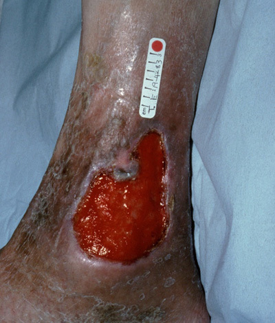
Leg Ulcers
Leg ulceration has commonly been defined as ‘a loss of skin below the knee on the leg or foot which takes more than six weeks to heal’1.
The majority of leg ulcers are caused by Venous Disease (70%), the remainder by either peripheral Arterial Disease (20%) or as a result of mixed aetiology, skin malignancies, metabolic disorders such as Diabetes, rheumatoid disorders, dermatological conditions or are self inflicted (10%). Most elderly patients who present are mulitfactorial2,3.
Appropriate management of the leg ulcer is dependent on a holistic assessment of the patient, the limb and the ulcer. Medical and surgical histories should be reviewed and both intrinsic and extrinsic risk factors considered. The aetiology of the ulcer should then be determined by using a Doppler and the ABPI (Ankle / Brachial Pressure Index) calculated4.
The ABPI detects arterial insufficiency. False readings can occur in patients with Diabetes or Atherosclerosis. This assessment should always be conducted by a trained healthcare professional, an inaccurate assessment and diagnosis could be detrimental for the patient5.
On assessment; venous and arterial ulcers often present differently in appearance. Venous Ulcers may be: Shallow in appearance and situated around the ‘gaiter region’ of the leg, oedematous, there may be ankle flare and hyper pigmentation (brown staining) and the leg may be ‘hardened‘. Arterial Ulcers may present as being: ‘punched out’ in appearance, poorly perfused, the legs and feet are cool to touch and there may be gangrene to the toes, the leg is often shiny, hairless and the skin may be tight2.

Leg ulcers should be managed locally with dressings and skin barrier preparations which will protect the peri-wound area from excoriation by the exudate. Dressings should be selected which manage the presenting conditions at the wound bed and optimise patient comfort. The wound should be assessed for the presence of infection, however many ulcers are colonised by microorganisms due to their chronicity. Many ulcers are malodorous due to the presence of the microorganisms, which have colonised the ulcer bed, in particularly bacterial species such as Pseudomonas aureginosa.
Leg ulcers, if they are venous in aetiology may be treated with compression therapy; multi-layer / short stretch bandaging and / or hosiery. The key is to ensure clinicians have the knowledge and experience to apply the bandages correctly. Once the ulcer is healed the patient should continue in compression hosiery to prevent reoccurrence2.
References:
- Dale, J.J., Callam, M.J., Ruckley, C.V., Harper, D.R., Berrey, P.N. (1983) Chronic ulcers of the leg: a study of prevalence in a Scottish community. Health Bull Edinburgh: 41; 6, 310 – 314
- Royal College of Nursing (1998) The management of patients with venous leg ulcers. RCN Institute.
- Moffatt, C. (2001) Leg Ulcers. In: Murray, S. (Ed) Vascular Disease: Nursing and Management. Whurr, London, 200 – 237.
- Vowden, P. and Vowden, K. (2002) Investigations in the management of lower limb ulceration. In: White, R. and Harding, K. (2002) Trends in Wound Care. Mark Allen Publishing Ltd.
Caveat: The information given is a guide only and should not replace clinical judgement.
