
Our guides and resources provide information and education to help patients succeed before and after surgery. We also provide educational support to healthcare professionals to develop clinical knowledge.
Guides and Educational Resources
Filter
Clinical Case Study
Breaking Free From Overactive Bladder
This case study explores the real-life experiences of five patients who successfully used Tensi+ to manage overactive bladder (OAB) symptoms. Their stories illustrate how this non-invasive, self-administered therapy, designed for use at home, helped reduce urgency and frequency, improve sleep, and restore confidence and independence.
E-Learning
HCP
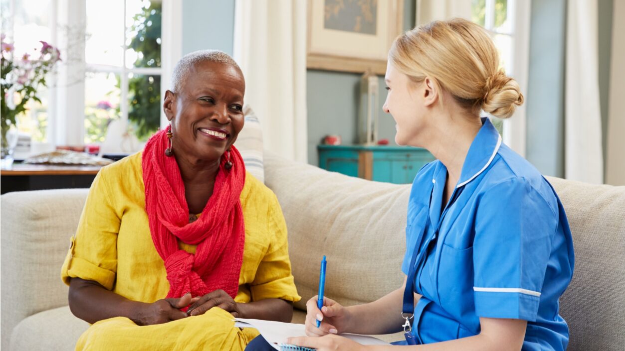
The Importance of Continence Care
This CPD-accredited course explores the role of fixation devices, also including urinary sheaths within continence care, focusing on how they enhance patient comfort and ensure stability, including the secure attachment of drainage bags.
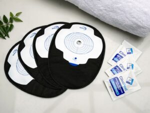
Clinical Case Study
HCP
Improving Sleep and Safety With a Urinary Sheath
This case study, written by Community Specialist Nurse Anna Moseley, explores the use of the CliniSure urinary sheath system in a patient with Parkinson’s disease and multiple comorbidities, highlighting how a tailored continence solution helped reduce fall risk, improve sleep, and restore quality of life for both patient and caregiver.
Guide
Understanding Catheterisation
An easy to understand guide to urinary catheterisation including what to expect from the procedure and how to maintain your catheter afterwards.
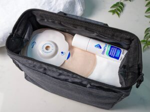
Clinical Case Study
HCP
Empowering Patient Choice in Intermittent Self-Catheterisation
Community Nurse Specialist Louise Harrison shares a case study highlighting the importance of patient choice when it comes to intermittent self-catheterisation (ISC) and how Curan Lady yielded positive results for a patient who had been experiencing challenges with ISC.
Clinical Case Study
HCP
Improving ISC Success for BPH Patients
This case study authored by Community Nurse Specialist Louise Harrison, reviews Curan Advantage with Tiemann Tip for a patient with benign prostatic hyperplasia addressing issues with insertion and improving confidence in managing to ISC independently.
Guide
Mental Health Guide for People with Incontinence
Designed to support people with continence issues, this guide offers practical advice and strategies to manage both the physical and mental health aspects of incontinence.
E-Learning
HCP
Fundamentals of Intermittent Self-Catheterisation
For patients who are unable to empty their bladder, intermittent self-catheterisation can have many benefits. Learn what ISC is, when it should or shouldn’t be used, and how to confidently support patients who are using it with this CPD-accredited course.
Guide
Nutrition Guide for Continence Patients
Written with dietitian Ruth Kander, this guide offers advice on beneficial foods, hydration, and lifestyle choices. It suggests what to include and avoid in your diet to support bladder health, reduce irritation, and manage incontinence. With practical meal ideas and tips, this guide helps you make bladder-friendly changes to your daily routine and help manage incontinence.
Guide
Women’s Pelvic Floor Exercise Guide
The female pelvic floor exercise guide developed in collaboration with pelvic health physiotherapist Natalia Vasquez provides practical exercises and routines tailored for women of all ages.
Guide
Urinary Incontinence Catheter Care Guide
This guide will help answer some of the questions you may have and give you tips and advice for how you can live your life with the catheter or continence appliance you've been advised to use. Whether you're looking for understanding, helpful tips, or solutions to common challenges, get the knowledge and confidence to navigate urinary incontinence and catheter use effectively with this free guide.
Guide
Sexual Wellbeing Continence Guide
The Sexual Wellbeing Continence Guide by CliniMed is designed to support individuals experiencing continence issues and their partners. The guide addresses the stigma, social isolation, and impact on intimacy associated with continence problems, offering practical advice on how to manage these issues while maintaining sexual wellbeing and healthy relationships.